


 2 / 8
2 / 8

2
( 8 5 5 ) 3 6 0 - G A M C
H Q
•
S P R I N G 2 0 1 5
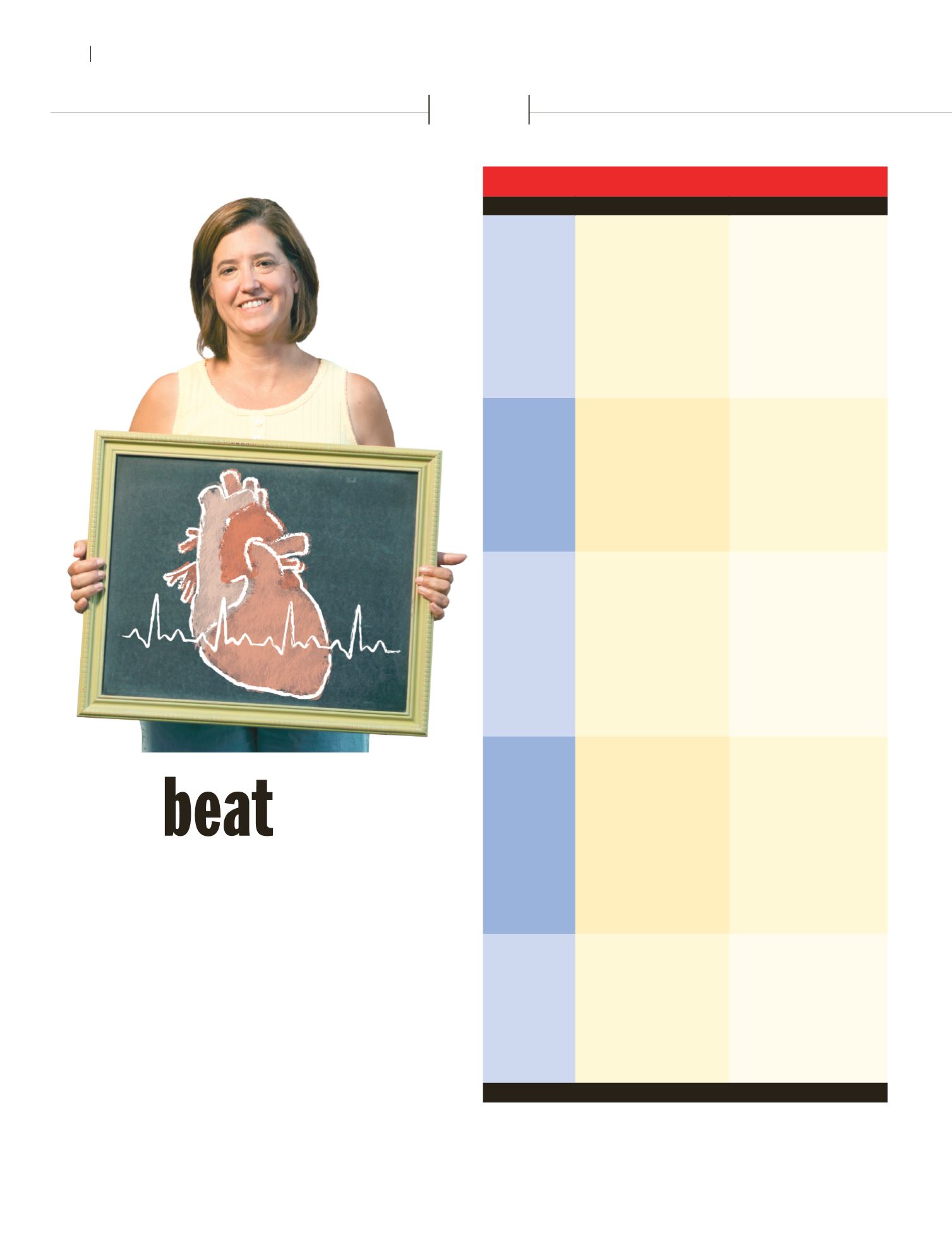
5 tests that detect heart disease
THE TEST
HOW IT’S DONE
WHY IT’S DONE
Echocardiography This test uses sound waves and
their echoes to make moving
pictures of your heart—much
like the ultrasound exams many
women have when pregnant. In
most cases, the sound waves
are sent from a handheld device
placed over your chest.
Your doctor might need infor-
mation about the size, shape
and function of your heart. For
example, the test can show how
well your heart is pumping. So it
might be used if you have signs
or symptoms of heart failure. Or
a doctor might want to know,
among other things, if your heart’s
valves are working properly or if
your heart is thick or enlarged.
Electrocardiogram
(EKG or ECG)
Up to 12 electrodes (soft, sticky
pads) are placed on the skin of
your chest, arms and legs. They
record your heart’s electrical
signals while you rest. Or, since
some heart problems occur only
at certain times, you might wear
a portable version of this device,
called a Holter or event monitor,
while you go about your day.
Your doctor may use it to help de-
tect problems ranging from dam-
age caused by past heart attacks
to an arrhythmia, in which your
heartbeat is too fast, too slow or
irregular. A pounding or fluttering
heartbeat are some signs that
may suggest a problem with your
heart’s rhythm or rate.
Stress test
Your heart is checked while
you exercise on a treadmill or
stationary bike. This gets your
heart working harder. If you
can’t exercise, you may be given
a medicine that makes your
heart beat as though you were
exercising.
Some problems are easier to
detect when your heart is working
harder. For one, during exercise,
clogged arteries may not be able
to meet the heart’s increased
need for oxygen-rich blood. That’s
why, for example, your doctor
might suggest a stress test to
learn why you have chest pain or
shortness of breath when you do
physical activities.
Cardiac
catheterization
For this procedure, your doctor
inserts a thin bendable tube
(catheter) into a blood vessel in
your arm or groin. Next, he or she
guides this catheter to an artery
in your heart. All of this typically
takes place in what’s called the
hospital’s cath lab.
Cardiac catheterization is com-
monly used to evaluate chest
pain and to set the stage for its
treatments. Once the catheter is
in place, your doctor can perform
x-ray tests to look for narrowed
heart arteries. He or she can even
treat those arteries through the
catheter by inflating a tiny balloon
at the site of the blockage. This
widens the artery, allowing more
blood to reach your heart.
Coronary
angiography
During cardiac catheterization,
your doctor may decide to take
x-ray pictures of the inside of
your heart. Through the catheter,
the doctor injects a dye that
travels through your bloodstream
to the coronary arteries, which
supply blood to your heart. The
dye makes the inside of these
arteries show up on the x-rays.
Angiography can show if fatty
buildup is clogging your heart
arteries. This can cause a heart
attack or chest discomfort called
angina. It may also be needed to
follow up on results from one of
the other cardiac tests.
Sources: American Heart Association; National Heart, Lung, and Blood Institute
1
2
3
4
5
hat’s troubling your ticker?
Often, that crucial question lies at
the heart of why your doctor might
want you to have certain cardiac
tests—especially if you’re having
chest pain, shortness of breath or
other symptoms of a possible heart
problem.
✦
“A variety of tests can
reveal why your heart may not be
functioning as well as it should be,”
explains Vahe Badalian, MD,
cardiologist at Glendale Adventist
Medical Center. This chart explains
some common ones.
The
goes on
GAMC offers a full range of cardiac tests and
screenings. Call the Heart & Vascular Institute
at (818) 863-4099 to learn more.
F